Tiny Echogenic Foci In Kidney
Nov 04, 2009 In order to be sure that a cyst is truly a benign fluid filled cyst, it is best to be at least 10 mm or 1 cm in diameter. Even small tumors that are less than 1 cm are most likely benign. As this grows with time, one will be able to tell benign cyst from solid tumor.
I have had horrible pain to the point that it has made me sick and cannot function or work, this has been going on since oct of 2012, but became progressively worse over the last week and a half. Pain in my right kidney that radiates to the front, nausea and stomach pain. Oct 2012 CT showed a 1-2 mm object in the upper right kidney. It has doubled since then and US revealed a 4mm echogenic focus with shadow. I also suffer from endometriosis and they are telling me that this is actually endo and advising me to have urgent laparoscopy to investigate and remove this object, any thoughts are welcome, Stephanie This discussion is related to.
The Content on this Site is presented in a summary fashion, and is intended to be used for educational and entertainment purposes only. It is not intended to be and should not be interpreted as medical advice or a diagnosis of any health or fitness problem, condition or disease; or a recommendation for a specific test, doctor, care provider, procedure, treatment plan, product, or course of action.
MedHelp is not a medical or healthcare provider and your use of this Site does not create a doctor / patient relationship. We disclaim all responsibility for the professional qualifications and licensing of, and services provided by, any physician or other health providers posting on or otherwise referred to on this Site and/or any Third Party Site. Never disregard the medical advice of your physician or health professional, or delay in seeking such advice, because of something you read on this Site. We offer this Site AS IS and without any warranties.
By using this Site you agree to the following. If you think you may have a medical emergency, call your physician or 911 immediately.
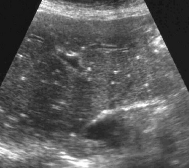
The calcifications in the renal sinus are often due to renal calculi and occasionally due to gas. Calcifications of the branches of the renal artery coursing through the sinus may be mistaken for renal calculi in some rare instances, resulting in a misdiagnosis.All hyperechoic or echogenic foci in the renal medulla are not caused by stones. Renal stones and gas are the commonly reported echogenic foci in the sinus region. However, in rare cases, the calcified vessel walls of renal arterial branches coursing through the sinus and renal parenchyma may be mistaken for renal calculi.
Songbringer review. A procedurally generated action game that begs to be played over and over and over. Songbringer review. Songbringer for PC game reviews & Metacritic score: Explore 308 million procedurally-generated worlds. Awaken and face dormant evil. Uncover lost technology and create powerful artifacts. Those familiar with the. Songbringer is the story of intergalactic explorer and top hat wearer Roq, who crash lands on the ever-adapting planet of Ekzera, separating him from his mothership, the titular Songbringer.
We report herein one such observation.A middle-aged woman diagnosed with diabetic nephropathy and hypertension presented with symptoms of urinary tract infection. Ultrasound of the abdomen revealed multiple calcifications at the junctional region of the sinus echoes and the medulla in both the kidneys. Although the calcifications were initially interpreted as renal stones, in view of their unconventional anatomical location, the patient was further subjected to Doppler ultrasonography and plain computed tomography which revealed the presence of calcification in the segmental and interlobar arteries, which was mistaken to be stones. During their course through the sinomedullary region, calcified branches of the renal artery, such as the segmental, interlobar, and arcuate arteries, may mimic nephrolithiasis in ultrasonographic imaging. In such a situation, a Doppler study is helpful in demonstrating the actual origin of calcification from the vessel wall to be distinct from the curvilinear appearance of calculi. A noncontrast CT study will also help to confirm the exact origin of calcification when it is observed in renal artery branches.